
Main menu

  • Home
  • Content
    • Current
    • Ahead of print
    • Archive
  • Info for
    • Authors
    • Reviewers
  • About Us
    • About Us
    • Conflict of Interest
    • Informed Consent
    • Human and Animal Rights
  • More
    • Alerts
    • Feedback
    • Folders
  • ascls.org
    • ascls.org

User menu

  • My alerts
  • Log in
  • My Cart

Search

  • Advanced search
American Society for Clinical Laboratory Science
  • ascls.org
    • ascls.org
  • My alerts
  • Log in
  • My Cart
American Society for Clinical Laboratory Science

Advanced Search

  • Home
  • Content
    • Current
    • Ahead of print
    • Archive
  • Info for
    • Authors
    • Reviewers
  • About Us
    • About Us
    • Conflict of Interest
    • Informed Consent
    • Human and Animal Rights
  • More
    • Alerts
    • Feedback
    • Folders
  • Follow ASCLS on Twitter
  • Visit ASCLS on Facebook
  • Follow ASCLS on Instagram
  • RSS Feed
Research ArticleClinical Practice

Laboratory Diagnosis of the Lupus Anticoagulant

Larry J. Smith
American Society for Clinical Laboratory Science January 2017, 30 (1) 7-14; DOI: https://doi.org/10.29074/ascls.30.1.7
Larry J. Smith
Abbott Diagnostics Division – Hematology Unit, Santa Clara, CA
PhD, SH(ASCP), HCLD/CC(ABB)
  • Find this author on Google Scholar
  • Find this author on PubMed
  • Search for this author on this site
  • Article
  • Figures & Data
  • Info & Metrics
  • PDF
Loading

Article Figures & Data

Figures

  • Figure 1.
    • Download figure
    • Open in new tab
    • Download powerpoint
    Figure 1.

    β2GPI and antibody. β2GPI and antibody to β2GPI binding to a phospholipid surface. B2GPI consists of 5 homologous domains. Domain V binds to the anionic phospholipid surface. The anti-β2GPI antibody binds to domain I.
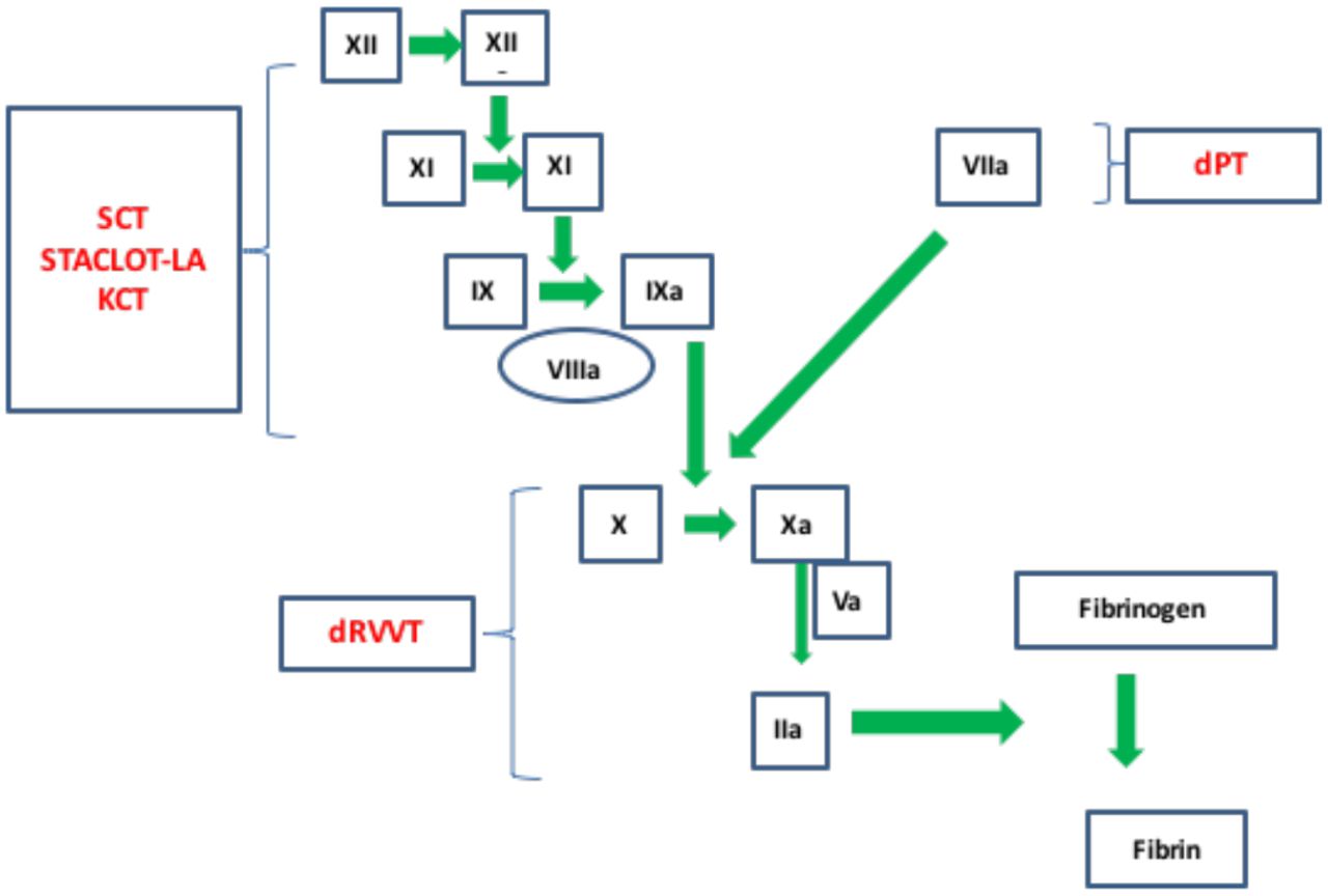
  • Figure 2.
    • Download figure
    • Open in new tab
    • Download powerpoint
    Figure 2.

    Interaction of specific LAC assays within the coagulation cascade. The heterogeneous nature of the LAC requires multiple assays to improve diagnostic sensitivity. (dPT, dilute Prothrombin Time; dRVVT, dilute Russell Viper Venom Time, KCT, Kaolin Clotting Time; SCT, Silica Clotting Time).
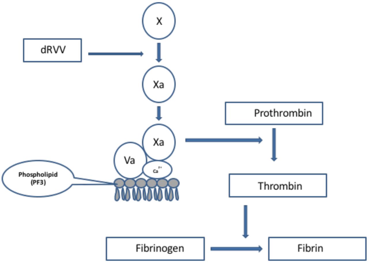
  • Figure 3.
    • Download figure
    • Open in new tab
    • Download powerpoint
    Figure 3.

    Mechanism of action of dRVV on the coagulation cascade. The enzyme present in Russell viper venom converts FX to FXa. FXa in the presence of FVa, calcium ions and phospholipids converts prothrombin to thrombin. Thrombin converts fibrinogen to fibrin.
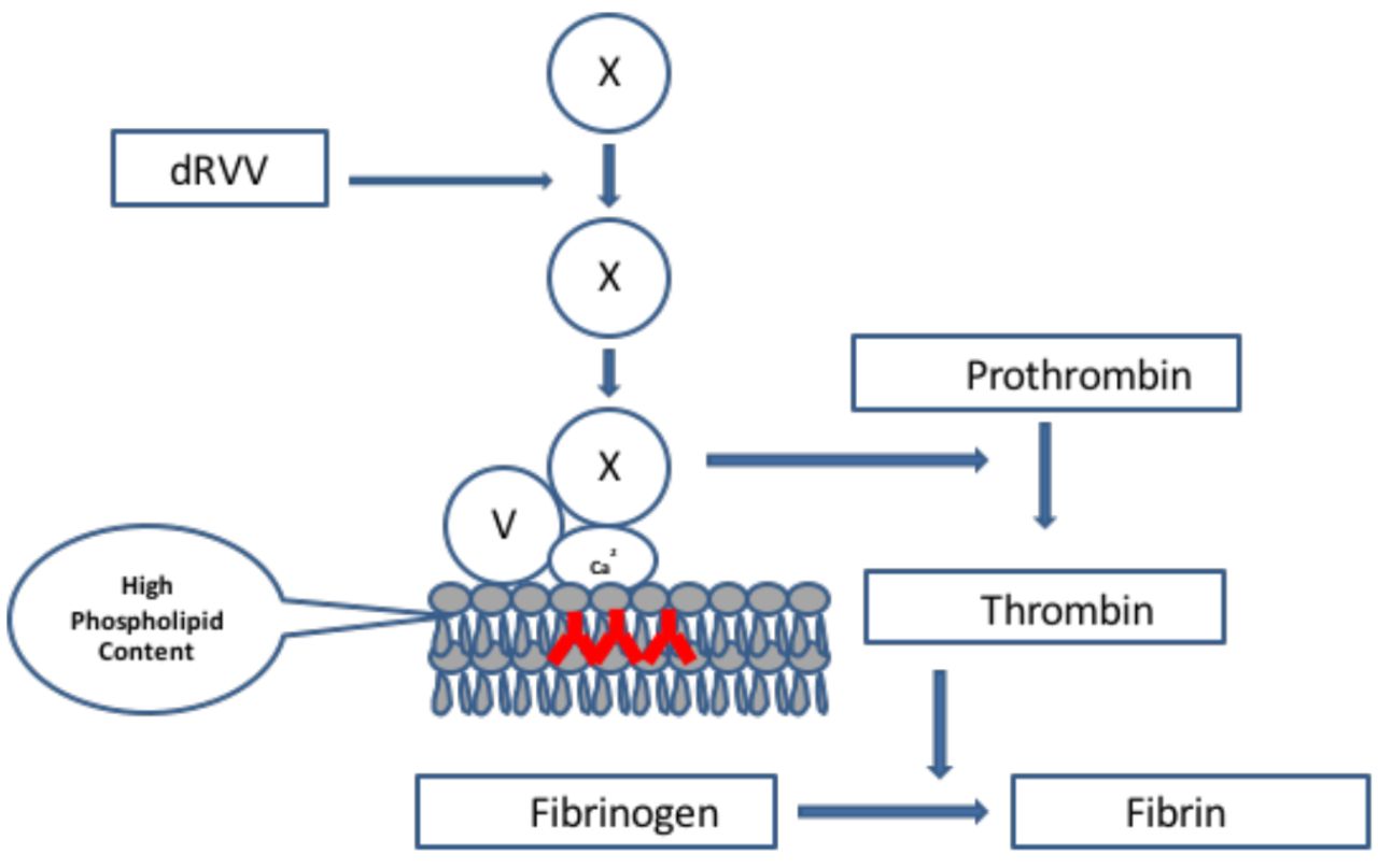
  • Figure 4.
    • Download figure
    • Open in new tab
    • Download powerpoint
    Figure 4.

    DRVVT screening assay. The screening assay requires a lupus sensitive reagent that contains a decreased amount of phospholipid. If a LAC is present, it will interfere with the prothrombinase complex binding to the phospholipid surface of platelets.

  • Figure 5.
    • Download figure
    • Open in new tab
    • Download powerpoint
    Figure 5.

    DRVVT confirm assay. The confirm assay requires a reagent that contains an increased concentration of phospholipid. If a LAC is present, it will neutralize the antibody facilitating binding of the prothrombinase complex onto the platelet phospholipid surface.
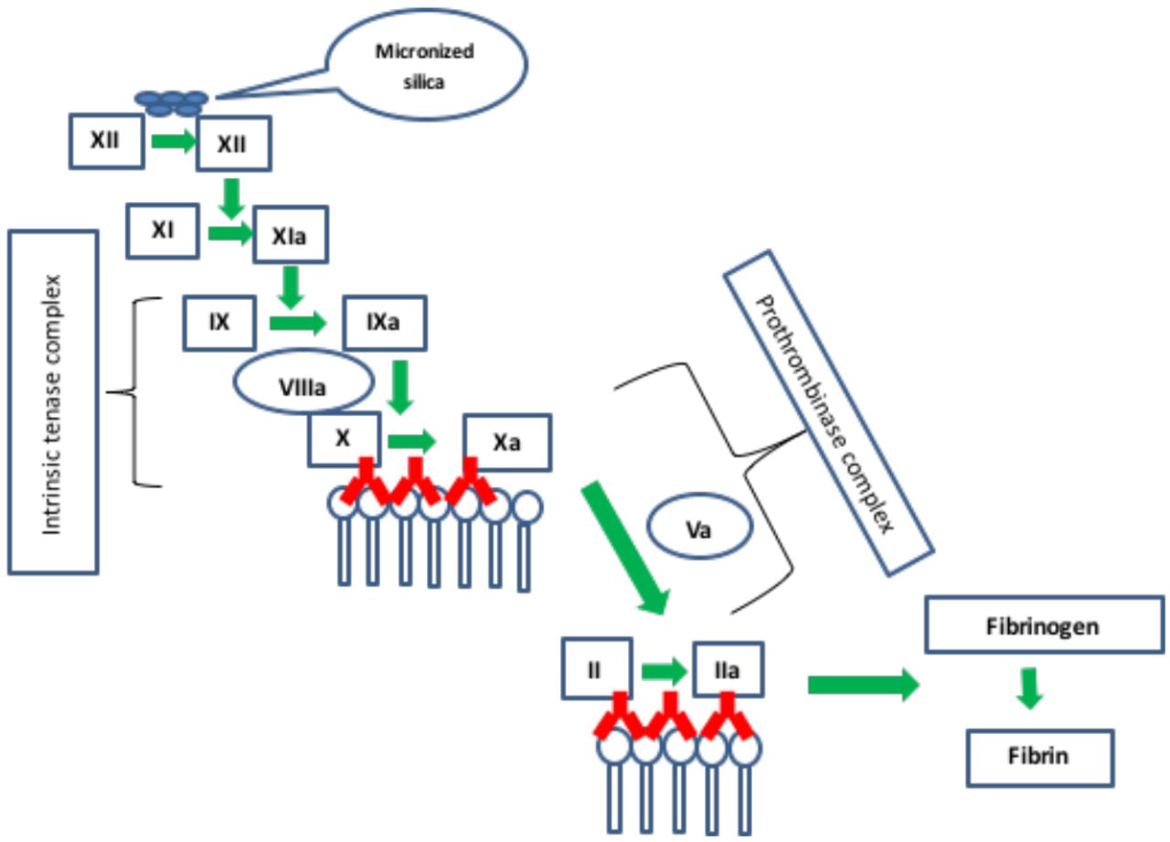
  • Figure 6.
    • Download figure
    • Open in new tab
    • Download powerpoint
    Figure 6.

    Silica Clotting Time assay (SCT). Micronized silica is used to activate the intrinsic pathway. LAC antibodies (in red) interfere with binding of the intrinsic tenase and prothrombinase complexes on the platelet surface.
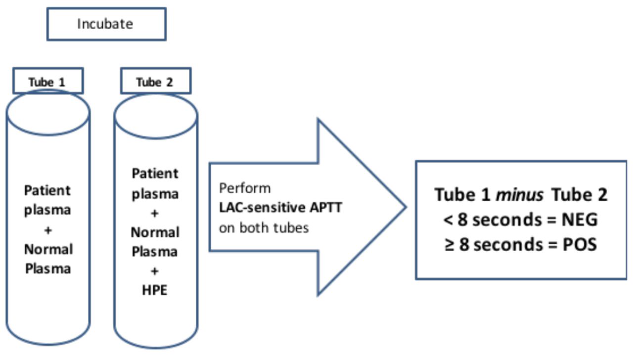
  • Figure 7.
    • Download figure
    • Open in new tab
    • Download powerpoint
    Figure 7.

    Hexagonal Phospholipid Neutralization assay (STACLOT-LA). Patient plasma plus normal plasma are incubated in the absence of HPE tube 1) and in the presence HPE (tube 2). An APTT is performed using a LAC sensitive reagent. The clotting time of tube 1 is compared to tube 2. If a LAC is present, tube 1 minus tube 2 should be ≥ to the established cut-off.

PreviousNext
Back to top

In this issue

American Society for Clinical Laboratory Science: 30 (1)
American Society for Clinical Laboratory Science
Vol. 30, Issue 1
Winter 2017
  • Table of Contents
  • Table of Contents (PDF)
  • Index by author
  • Front Matter (PDF)
Print
Download PDF
Article Alerts
Sign In to Email Alerts with your Email Address
Email Article

Thank you for your interest in spreading the word on American Society for Clinical Laboratory Science.

NOTE: We only request your email address so that the person you are recommending the page to knows that you wanted them to see it, and that it is not junk mail. We do not capture any email address.

Enter multiple addresses on separate lines or separate them with commas.
Laboratory Diagnosis of the Lupus Anticoagulant
(Your Name) has sent you a message from American Society for Clinical Laboratory Science
(Your Name) thought you would like to see the American Society for Clinical Laboratory Science web site.
CAPTCHA
This question is for testing whether or not you are a human visitor and to prevent automated spam submissions.
Citation Tools
Laboratory Diagnosis of the Lupus Anticoagulant
Larry J. Smith
American Society for Clinical Laboratory Science Jan 2017, 30 (1) 7-14; DOI: 10.29074/ascls.30.1.7

Citation Manager Formats

  • BibTeX
  • Bookends
  • EasyBib
  • EndNote (tagged)
  • EndNote 8 (xml)
  • Medlars
  • Mendeley
  • Papers
  • RefWorks Tagged
  • Ref Manager
  • RIS
  • Zotero
Request Permissions
Share
Laboratory Diagnosis of the Lupus Anticoagulant
Larry J. Smith
American Society for Clinical Laboratory Science Jan 2017, 30 (1) 7-14; DOI: 10.29074/ascls.30.1.7
Twitter logo Facebook logo Mendeley logo
  • Tweet Widget
  • Facebook Like
  • Google Plus One

Jump to section

  • Article
    • Abstract
    • INTRODUCTION
    • CONCLUSION
    • REFERENCES
  • Figures & Data
  • Info & Metrics
  • PDF

Related Articles

  • No related articles found.
  • Google Scholar

Cited By...

  • No citing articles found.
  • Google Scholar

More in this TOC Section

  • Utility of Immunofixation as a Follow-up to Select Abnormal Serum Protein Electrophoresis Patterns and Suggestions for Clinical Correlation
  • Public Datasets: A Foundation to Artificial Intelligence in Health Care
  • Comparison of Laboratory Scientist and CellaVision DM9600 Classification of Reactive Lymphocytes
Show more Clinical Practice

Similar Articles

Keywords

  • Antiphospholipid antibody
  • anticardiolipin antibody
  • beta-2 glycoprotein I antibody
  • lupus anticoagulant
  • dilute Russell's viper venom time
  • silica clotting time
  • STACLOT-LA

© 2025 The American Society for Clinical Laboratory Science

Powered by HighWire