
Main menu

  • Home
  • Content
    • Current
    • Ahead of print
    • Archive
  • Info for
    • Authors
    • Reviewers
  • About Us
    • About Us
    • Conflict of Interest
    • Informed Consent
    • Human and Animal Rights
  • More
    • Alerts
    • Feedback
    • Folders
  • ascls.org
    • ascls.org

User menu

  • My alerts
  • Log in
  • My Cart

Search

  • Advanced search
American Society for Clinical Laboratory Science
  • ascls.org
    • ascls.org
  • My alerts
  • Log in
  • My Cart
American Society for Clinical Laboratory Science

Advanced Search

  • Home
  • Content
    • Current
    • Ahead of print
    • Archive
  • Info for
    • Authors
    • Reviewers
  • About Us
    • About Us
    • Conflict of Interest
    • Informed Consent
    • Human and Animal Rights
  • More
    • Alerts
    • Feedback
    • Folders
  • Follow ASCLS on Twitter
  • Visit ASCLS on Facebook
  • Follow ASCLS on Instagram
  • RSS Feed
Research ArticleResearch and Reports

Next-generation Sequencing for Outbreak Investigation in the Clinical Microbiology Laboratory

Caitlin E. Dougherty and Erin Graf
American Society for Clinical Laboratory Science April 2019, 32 (2) 70-77; DOI: https://doi.org/10.29074/ascls.2019001750
Caitlin E. Dougherty
Children’s Hospital of Philadelphia
  • Find this author on Google Scholar
  • Find this author on PubMed
  • Search for this author on this site
Erin Graf
Children’s Hospital of Philadelphia
  • Find this author on Google Scholar
  • Find this author on PubMed
  • Search for this author on this site
  • Article
  • Figures & Data
  • Info & Metrics
  • PDF
Loading

Article Figures & Data

Figures

  • Figure 1.
    • Download figure
    • Open in new tab
    • Download powerpoint
    Figure 1.

    Conventional and NGS methods for viral outbreak investigation. The Sanger workflow (A) involves amplification of 1 or several regions of the genome with primers (blue arrows), analysis via capillary electrophoresis (multicolor peaks), and comparison of generally partial genome sequence between samples to infer relatedness. mNGS workflow (B) involves preparation of libraries (either RNA- or DNA-dependent on virus of interest) that include all RNA or DNA in the clinical sample (including mostly human and commensal flora). Reads can be aligned to a reference viral genome as pictured on the left. Alternatively, as pictured on the right, nonviral reads can be removed or viral reads can be binned via bioinformatic pipelines, and the reads of interest can be strung together via overlapping regions to create a de novo assembly. Either way, the end goal is to generate a complete viral genome with sufficient coverage of reads across the entire sequence length for comparison between samples.
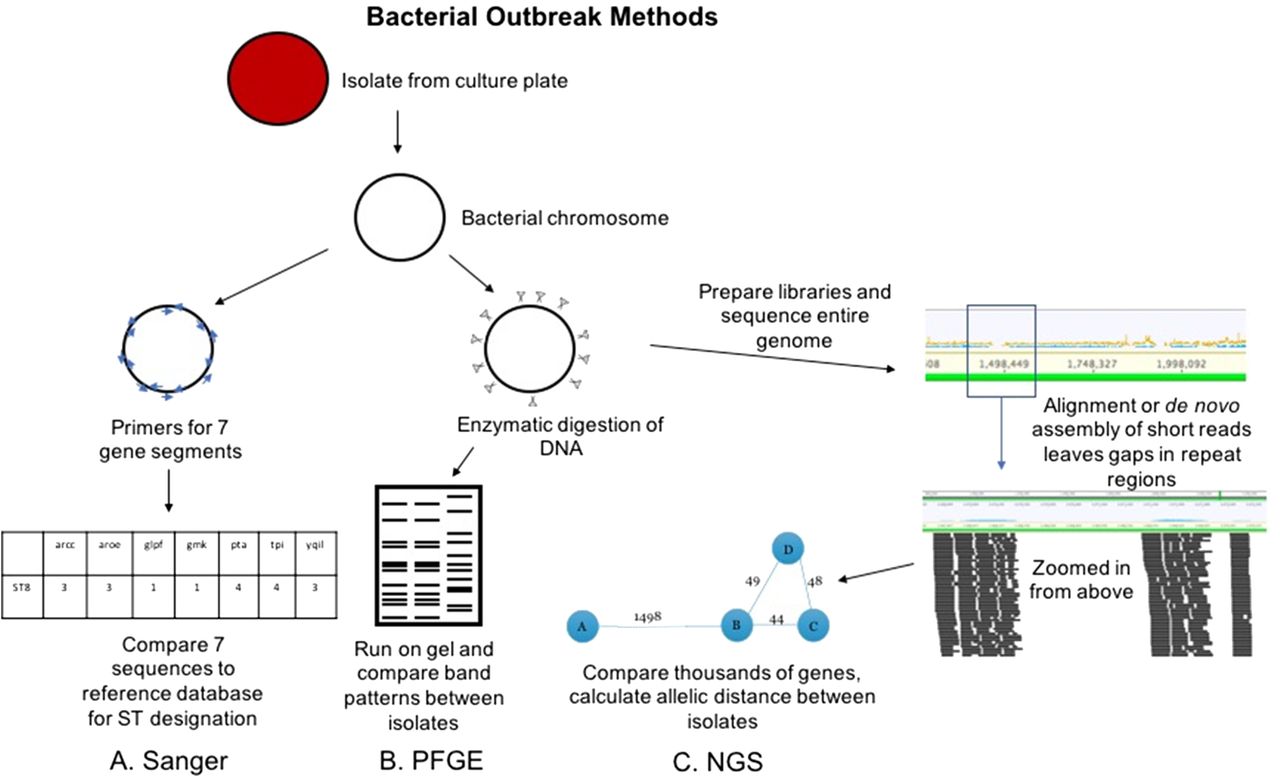
  • Figure 2.
    • Download figure
    • Open in new tab
    • Download powerpoint
    Figure 2.

    Conventional and NGS methods for bacterial outbreak investigation. The Sanger workflow (A) involves amplification of 7 gene regions with primers (blue arrows), analysis via capillary electrophoresis (like Figure 1), and comparison of the sequences to known sequence types (STs). PFGE workflow (B) involves enzymatic cutting of the bacterial chromosome followed by gel electrophoresis that alternates pulses of current in various directions to separate DNA fragments at higher resolution. The band patterns are compared between isolates to determine relatedness. NGS workflow (C) involves preparation of libraries from fragmented chromosomal DNA. Reads are assembled (either aligned or de novo as in Figure 1) to form contiguous sequences (contigs). From these contigs, thousands of genes or alleles are compared for similarity between isolates. An allelic distance, or number of alleles that are different between isolates, is calculated, and this can be visualized as the bubble and line plot shown above.

  • Figure 3.
    • Download figure
    • Open in new tab
    • Download powerpoint
    Figure 3.

    Normalized adenoviral reads are higher for samples with less background nucleic acid when adjusted for viral titer. The percent of adenoviral reads normalized to the total number of NGS reads is displayed on the x-axis. The adenovirus crossing cycle threshold (surrogate for viral titer) by real-time PCR is displayed on the y-axis. Samples with lower background human nucleic acid are in red and blue (eye and environmental samples, respectively), whereas samples with high human background are in green (respiratory samples). Mean CT -adjusted percent AdV per total reads was 5.87 times higher for eye specimens compared with respiratory specimens and was significantly different by ANOCOVA analysis (p = 0.017). CT, real-time PCR crossing cycle threshold.

PreviousNext
Back to top

In this issue

American Society for Clinical Laboratory Science: 32 (2)
American Society for Clinical Laboratory Science
Vol. 32, Issue 2
1 Apr 2019
  • Table of Contents
  • Index by author
Print
Download PDF
Article Alerts
Sign In to Email Alerts with your Email Address
Email Article

Thank you for your interest in spreading the word on American Society for Clinical Laboratory Science.

NOTE: We only request your email address so that the person you are recommending the page to knows that you wanted them to see it, and that it is not junk mail. We do not capture any email address.

Enter multiple addresses on separate lines or separate them with commas.
Next-generation Sequencing for Outbreak Investigation in the Clinical Microbiology Laboratory
(Your Name) has sent you a message from American Society for Clinical Laboratory Science
(Your Name) thought you would like to see the American Society for Clinical Laboratory Science web site.
CAPTCHA
This question is for testing whether or not you are a human visitor and to prevent automated spam submissions.
Citation Tools
Next-generation Sequencing for Outbreak Investigation in the Clinical Microbiology Laboratory
Caitlin E. Dougherty, Erin Graf
American Society for Clinical Laboratory Science Apr 2019, 32 (2) 70-77; DOI: 10.29074/ascls.2019001750

Citation Manager Formats

  • BibTeX
  • Bookends
  • EasyBib
  • EndNote (tagged)
  • EndNote 8 (xml)
  • Medlars
  • Mendeley
  • Papers
  • RefWorks Tagged
  • Ref Manager
  • RIS
  • Zotero
Request Permissions
Share
Next-generation Sequencing for Outbreak Investigation in the Clinical Microbiology Laboratory
Caitlin E. Dougherty, Erin Graf
American Society for Clinical Laboratory Science Apr 2019, 32 (2) 70-77; DOI: 10.29074/ascls.2019001750
Twitter logo Facebook logo Mendeley logo
  • Tweet Widget
  • Facebook Like
  • Google Plus One

Jump to section

  • Article
    • LEARNING OBJECTIVES
    • ABSTRACT
    • INTRODUCTION
    • HOW DOES NGS WORK FOR WHOLE VIRAL GENOME SEQUENCING?
    • CLINICAL APPLICATIONS OF NGS FOR VIRAL OUTBREAK INVESTIGATIONS
    • HOW DOES NGS WORK FOR WHOLE BACTERIAL GENOME SEQUENCING?
    • CLINICAL APPLICATIONS OF NGS FOR BACTERIAL OUTBREAK INVESTIGATIONS
    • OTHER CONSIDERATIONS FOR BACTERIAL WGS
    • WORKFORCE NEEDS FOR NGS IN OUTBREAK INVESTIGATION
    • SUMMARY
    • References
  • Figures & Data
  • Info & Metrics
  • PDF

Related Articles

  • No related articles found.
  • Google Scholar

Cited By...

  • No citing articles found.
  • Google Scholar

More in this TOC Section

  • Barriers to Implementation of Quality Management Systems in Laboratories: Lessons from the Southern Africa TB Health Systems Project
  • Polyagglutination: Lectin Isolation for T-Activated Red Cells
  • Strength of Direct Antiglobulin Test as an Indicator of Disease Severity in Autoimmune Hemolytic Anemia
Show more Research and Reports

Similar Articles

Keywords

  • cgMLST - core genome MLST
  • GUI - graphical user interface
  • MLST - multilocus sequence typing
  • mNGS - metagenomic NGS
  • MRSA - methicillin-resistant Staphylococcus aureus
  • NGS - next-generation sequencing
  • PCR - polymerase chain reaction
  • PFGE - pulsed field gel electrophoresis
  • SNV - single-nucleotide variation
  • ST - sequence type
  • WGS - whole-genome sequencing
  • whole-genome sequencing
  • outbreak investigation
  • next-generation sequencing
  • metagenomics

© 2025 The American Society for Clinical Laboratory Science

Powered by HighWire